The distinctive gurgling sounds emanating from a hernia can be both puzzling and concerning for patients. These acoustic manifestations occur when bowel contents move through herniated tissue, creating characteristic noises that range from gentle bubbling to more pronounced rumbling sounds. While gurgling hernias often represent normal bowel movement through the hernial sac, they can also signal potentially serious complications that require immediate medical attention.
Understanding the significance of these sounds is crucial for both patients and healthcare providers. The transition from normal gurgling to silent periods, accompanied by other warning signs, may indicate life-threatening complications such as bowel obstruction or strangulation. Early recognition of these danger signals can mean the difference between routine surgical repair and emergency intervention with significantly higher risks.
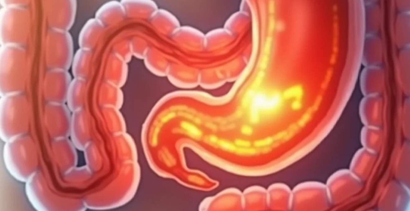
Understanding gurgling hernias: pathophysiology and acoustic manifestations
The pathophysiology behind gurgling hernias involves the protrusion of bowel segments through weakened areas in the abdominal wall. When intestinal contents move through these displaced bowel loops, they create the characteristic acoustic phenomena that patients describe as gurgling, bubbling, or rumbling sounds. This process is fundamentally similar to normal peristalsis, but the altered anatomical position and potential constriction at the hernial neck can modify the acoustic properties.
The intensity and frequency of gurgling sounds often correlate with several factors, including the size of the hernial defect, the amount of bowel involved, and the presence of any obstruction. Louder, more frequent gurgling typically indicates increased bowel activity , which may occur as the intestines attempt to push contents through a narrowed space. Conversely, diminished or absent sounds can suggest more serious complications.
Bowel movement through hernial sacs: peristaltic sound generation
Normal peristaltic waves continue to occur within herniated bowel segments, creating the acoustic manifestations patients notice. The hernial sac acts as a resonating chamber, potentially amplifying these sounds compared to intra-abdominal bowel sounds. The frequency and character of these sounds depend on the type of bowel involved, with small bowel typically producing higher-pitched, more frequent sounds than large bowel.
The mechanical properties of the hernial sac itself contribute to sound transmission. A thin-walled sac with minimal surrounding tissue allows for clearer sound transmission, while thick-walled or fibrotic sacs may muffle these acoustic signals. Understanding these variations helps clinicians interpret the clinical significance of different sound patterns .
Differentiation between reducible and incarcerated hernia sounds
Reducible hernias typically produce intermittent gurgling sounds that may vary with position and activity level. These sounds often become more pronounced during periods of increased intra-abdominal pressure, such as coughing, straining, or physical exertion. The ability to reduce the hernia manually usually correlates with continued, albeit variable, acoustic activity.
Incarcerated hernias present a more complex acoustic picture. Initially, sounds may become more prominent and constant as the bowel attempts to overcome the mechanical obstruction. However, as swelling and inflammation develop, the acoustic characteristics change. The progression from hyperactive to hypoactive bowel sounds within a hernia often signals increasing severity .
Anatomical locations where gurgling occurs: inguinal vs umbilical presentations
Inguinal hernias, being the most common type, frequently present with gurgling sounds that patients can localise to the groin area. The anatomical constraints of the inguinal canal can create unique acoustic properties, with sounds often radiating along the spermatic cord in males or round ligament pathway in females. The deeper location of some inguinal hernias may make sounds less audible externally.
Umbilical hernias typically produce more easily audible gurgling due to their superficial location and the thin-walled nature of the umbilical defect. Patients often describe being able to feel the bubbling sensation directly beneath their fingertips when palpating the area. The central location also means sounds may be mistaken for normal abdominal sounds, making clinical assessment crucial.
Strangulated hernia complications: silent periods and absent bowel sounds
The development of strangulation represents a surgical emergency where the blood supply to herniated bowel becomes compromised. One of the most ominous signs is the transition from active gurgling to periods of silence. This acoustic change reflects the bowel’s inability to maintain normal peristaltic activity due to ischaemia and subsequent paralysis of the intestinal smooth muscle.
The absence of previously present gurgling sounds in a known hernia should always be considered a potential sign of strangulation until proven otherwise through immediate medical evaluation.
Critical warning signs of hernia strangulation and obstruction
Recognition of warning signs that indicate progression from a simple gurgling hernia to a surgical emergency requires understanding both the acoustic changes and accompanying clinical manifestations. The evolution from benign gurgling to dangerous complications often follows a predictable pattern, though the timeline can vary significantly between patients. Early identification of these danger signals enables prompt intervention, potentially preventing life-threatening consequences such as bowel necrosis, perforation, and sepsis.
The clinical presentation of hernia complications involves multiple organ systems, reflecting both local effects of the hernia and systemic responses to bowel obstruction or ischaemia. Healthcare providers must maintain a high index of suspicion , as elderly patients or those with cognitive impairment may not clearly communicate symptom progression. The combination of multiple warning signs significantly increases the likelihood of serious complications requiring immediate surgical intervention.
Sudden cessation of gurgling: ischaemic bowel indicators
The abrupt cessation of gurgling sounds in a previously active hernia represents one of the most concerning acoustic changes. This silence often indicates that the herniated bowel segment has lost its ability to generate normal peristaltic waves due to compromised blood supply. The transition typically occurs over hours rather than days, making patient education about monitoring acoustic changes crucial.
Accompanying this acoustic silence, patients may experience intensifying pain that shifts from the cramping, intermittent discomfort associated with obstruction to a more constant, severe pain characteristic of ischaemia. The bowel’s attempt to maintain function despite compromised circulation initially leads to increased activity, but eventual failure results in the ominous quiet period that signals tissue death may be imminent.
Erythema and skin discolouration over hernial sites
Visual changes in the skin overlying a hernia provide important clues about the underlying pathological processes. Early inflammatory changes manifest as erythema or redness, indicating increased blood flow to the area as part of the inflammatory response. As complications progress, the skin may develop a dusky or purplish discolouration, suggesting venous congestion or more serious vascular compromise.
The progression of skin colour changes often correlates with the severity of internal complications. Pale or mottled skin over a hernia may indicate arterial insufficiency , while dark red or purple discolouration suggests venous obstruction or tissue death. These visual cues, combined with changes in acoustic activity, provide valuable diagnostic information about the urgency of the situation.
Nausea, vomiting, and bilious emesis patterns
Gastrointestinal symptoms accompany hernia complications as the body responds to bowel obstruction or ischaemia. Initial nausea may be mild and intermittent, but as complications progress, vomiting becomes more frequent and characteristic. The nature of the vomitus provides diagnostic clues, with clear or food-containing material initially progressing to bilious (green-yellow) content as small bowel obstruction develops.
In cases of high-grade small bowel obstruction, the vomiting becomes more projectile and frequent. Faeculent vomiting, though rare, represents a late and ominous sign indicating severe, prolonged obstruction with possible bacterial overgrowth. The combination of persistent vomiting with cessation of hernia gurgling sounds demands immediate medical evaluation.
Abdominal distension and absence of flatus passage
Progressive abdominal distension develops as bowel obstruction prevents the normal passage of gas and fluid through the intestinal tract. Patients often notice increasing abdominal girth and tightness, particularly in the supraumbilical region where small bowel distension is most prominent. The severity of distension correlates with the level and completeness of the obstruction.
The inability to pass flatus represents a significant clinical finding that, combined with other symptoms, strongly suggests bowel obstruction. Patients may report feeling the need to pass gas but being unable to do so, or they may notice a complete absence of normal bowel function. This symptom, particularly when coupled with cessation of hernia gurgling, indicates the need for urgent surgical evaluation.
Tachycardia and systemic inflammatory response syndrome signs
Systemic manifestations of hernia complications reflect the body’s response to stress, fluid losses, and potential infection. Tachycardia often represents the earliest cardiovascular sign, initially due to dehydration from vomiting and later from systemic inflammatory responses. Heart rate elevation above baseline, particularly when persistent, indicates significant physiological stress.
As complications progress, patients may develop fever, indicating either systemic inflammatory response syndrome or frank infection. The combination of tachycardia, fever, and altered mental status suggests severe complications requiring immediate intensive management. These systemic signs, when present with acoustic changes in hernia sounds, mandate emergency intervention.
Emergency assessment protocols for gurgling hernias
The systematic evaluation of patients presenting with gurgling hernias requires a structured approach that combines clinical assessment, diagnostic imaging, and laboratory investigations. Emergency departments must maintain protocols that enable rapid differentiation between benign hernias with normal gurgling and those requiring urgent surgical intervention. The assessment process should be time-sensitive, as delays in diagnosis can lead to irreversible complications.
Modern emergency assessment protocols emphasise the importance of serial examinations, as the clinical picture in hernia complications can evolve rapidly. A patient who appears stable initially may deteriorate within hours , making continuous monitoring and reassessment essential components of emergency care. The integration of clinical findings with advanced diagnostic modalities provides the most reliable approach to identifying patients requiring immediate surgical intervention.
Physical examination techniques: auscultation and palpation methods
Systematic physical examination begins with visual inspection of the hernia, noting size, shape, colour changes, and any obvious signs of distress. Auscultation should be performed with the patient in various positions, as gurgling sounds may be position-dependent. The stethoscope should be placed directly over the hernia and in surrounding areas to characterise the acoustic properties and localise the source of sounds.
Palpation techniques require gentle assessment of the hernia’s consistency, reducibility, and associated tenderness. Excessive manipulation should be avoided in cases where strangulation is suspected , as this may worsen the obstruction or compromise already tenuous blood supply. The examination should include assessment of pulse quality, skin temperature, and capillary refill in the affected area.
CT imaging protocols for suspected bowel obstruction
Computed tomography with intravenous contrast represents the gold standard for evaluating suspected hernia complications. The imaging protocol should include thin-section acquisition through the abdomen and pelvis, with particular attention to the hernial defect and contents. Oral contrast may be contraindicated in cases of high-grade obstruction or when surgery is imminent, but IV contrast remains essential for assessing bowel viability.
Key imaging findings include bowel wall thickening, fluid collection within the hernia sac, and signs of bowel obstruction such as proximal dilatation with distal decompression. The “whirl sign” on CT imaging may indicate bowel volvulus within the hernia , while poor enhancement of the bowel wall suggests ischaemia. These findings, combined with clinical assessment, guide surgical decision-making.
Laboratory markers: white cell count and lactate levels
Laboratory investigations provide supporting evidence for hernia complications, though normal values do not exclude the diagnosis. White blood cell count elevation indicates inflammatory response, but may be absent in elderly patients or those with compromised immune systems. The degree of leucocytosis often correlates with the severity of complications, with marked elevation suggesting possible bowel necrosis or perforation.
Serum lactate levels serve as a valuable marker of tissue hypoxia and anaerobic metabolism. Elevated lactate suggests significant bowel ischaemia and correlates with increased morbidity and mortality. Serial lactate measurements may be more informative than isolated values , as trending can indicate improving or deteriorating clinical status. Other markers such as C-reactive protein and procalcitonin may support the diagnosis but are less specific.
Differentiation from pseudo-obstruction and ileus presentations
Distinguishing true mechanical obstruction from functional disorders such as paralytic ileus or pseudo-obstruction can be challenging but is crucial for appropriate management. Pseudo-obstruction typically presents with more generalised abdominal distension and absent bowel sounds throughout the abdomen, rather than localised to the hernia. The clinical history often reveals precipitating factors such as medications, metabolic disorders, or recent surgery.
Imaging findings help differentiate these conditions, with mechanical obstruction showing a clear transition point at the hernial defect, while pseudo-obstruction demonstrates uniform bowel dilatation without a specific obstructing lesion. The presence of gurgling specifically localised to the hernia strongly suggests mechanical rather than functional obstruction . Treatment approaches differ significantly, making accurate differentiation essential.
Immediate medical intervention requirements and surgical timing
The decision for immediate surgical intervention in patients with gurgling hernias depends on multiple factors, including the presence of strangulation, complete bowel obstruction, and the patient’s overall physiological status. Emergency surgery is indicated when clinical findings suggest compromised bowel viability, as delays significantly increase morbidity and mortality rates. The surgical approach may range from simple hernia reduction and repair to bowel resection with anastomosis, depending on the degree of tissue damage encountered.
Preoperative optimisation must be balanced against the urgency of intervention. Patients with signs of strangulation require immediate surgery, while those with partial obstruction may benefit from brief periods of conservative management with careful monitoring. The window for successful conservative management is typically measured in hours rather than days . Fluid resuscitation, nasogastric decompression, and antibiotic prophylaxis form the cornerstone of immediate medical management while preparing for surgery.
Anaesthetic considerations in emergency hernia surgery include the increased risk of aspiration due to bowel obstruction and potential haemodynamic instability from fluid losses and systemic inflammation. Rapid sequence induction with cricoid pressure and appropriate positioning minimise aspiration risk. The surgical team must be prepared for potential bowel resection, with appropriate instruments and expertise available. Intraoperative findings determine the extent of intervention required, from simple reduction to complex bowel reconstruction.
Long-term complications of untreated gurgling hernias
The natural history of untreated gurgling hernias involves progressive enlargement of the hernial defect and increased risk of complications over time. Statistics indicate that approximately 10-15% of inguinal hernias will eventually develop complications if left untreated, with incarceration rates of 2-3% per year and strangulation occurring in 0.3-3% annually. These figures underscore the importance of timely surgical intervention, particularly in patients with symptomatic hernias.
Chronic intermittent incarceration can lead to adhesion formation between the herniated bowel and the surrounding tissues, making subsequent surgical repair more complex and increasing the risk of bowel injury during surgery. Repeated episodes of partial obstruction may cause bowel wall thickening and reduced peristaltic function , potentially leading to chronic digestive symptoms even after successful hernia repair. The psychological impact of living with an untreated hernia, including anxiety about potential complications and activity restrictions, significantly affects quality of life.
Patients who experience gurgling hernias face a 25% increased risk of developing complete bowel obstruction within five years compared to those with non-symptomatic hernias.
Long-term complications also include the development of chronic pain syndromes, particularly in cases where nerve entrapment occurs alongside bowel incarceration. The inflammatory response associated with chronic incarceration can involve nearby nerves, leading to persistent pain even after hernia reduction. Additionally, patients may develop compensatory movement patterns to avoid discomfort
, which can compound existing musculoskeletal issues and lead to secondary health problems affecting mobility and overall functional capacity.
Prevention strategies and conservative management approaches
The cornerstone of hernia prevention lies in addressing modifiable risk factors and implementing lifestyle modifications that reduce intra-abdominal pressure. Weight management through balanced nutrition and regular exercise helps decrease the mechanical stress on abdominal wall structures. Studies demonstrate that maintaining a body mass index below 25 kg/m² significantly reduces hernia recurrence rates following surgical repair. Dietary modifications should emphasise high-fibre foods to prevent constipation, which is a major contributing factor to increased abdominal pressure during defecation.
Proper lifting techniques form a crucial component of hernia prevention strategies. The principle of lifting with legs rather than the back, maintaining neutral spine alignment, and avoiding breath-holding during exertion helps distribute forces more effectively across the body. Workplace ergonomic assessments and training programmes have shown measurable success in reducing hernia incidence among manual labourers and those engaged in repetitive lifting activities.
Smoking cessation represents another vital preventive measure, as chronic coughing associated with tobacco use creates repeated spikes in intra-abdominal pressure. The cessation of smoking not only reduces the risk of developing new hernias but also improves healing outcomes following surgical repair. Former smokers show a 40% reduction in hernia recurrence rates compared to active smokers undergoing similar surgical procedures.
For patients with existing gurgling hernias who are not immediate surgical candidates, conservative management approaches can help minimise symptom progression and reduce complication risks. Dietary modifications include eating smaller, more frequent meals to reduce gastric distension and avoiding carbonated beverages that increase intestinal gas production. The timing of meals in relation to physical activity should also be considered, with patients advised to avoid strenuous activity immediately after eating.
Conservative management can effectively control symptoms in up to 60% of patients with small, reducible hernias, though surgical repair remains the definitive treatment for most cases.
Activity modification programmes help patients identify and avoid movements or positions that exacerbate hernia symptoms. This includes temporary restrictions on heavy lifting, prolonged standing, and high-impact activities that increase intra-abdominal pressure. Physical therapy interventions focusing on core strengthening and postural awareness can provide additional support, though these measures should complement rather than replace surgical consultation when indicated.
Abdominal binding or truss devices may provide temporary symptom relief in select patients, particularly those who are poor surgical candidates due to comorbidities. However, these devices should not be considered a permanent solution, as they do not address the underlying structural defect and may actually contribute to tissue weakness over time. The use of external supports requires careful monitoring to ensure they do not mask the development of complications such as strangulation.
Patient education remains perhaps the most critical component of conservative management. Individuals must understand the warning signs that necessitate immediate medical attention, including sudden increases in pain, changes in hernia appearance, cessation of gurgling sounds, or development of nausea and vomiting. Regular follow-up appointments allow healthcare providers to monitor hernia progression and adjust management strategies as needed, ensuring that the transition to surgical intervention occurs at the optimal time to minimise complication risks while maximising patient outcomes.