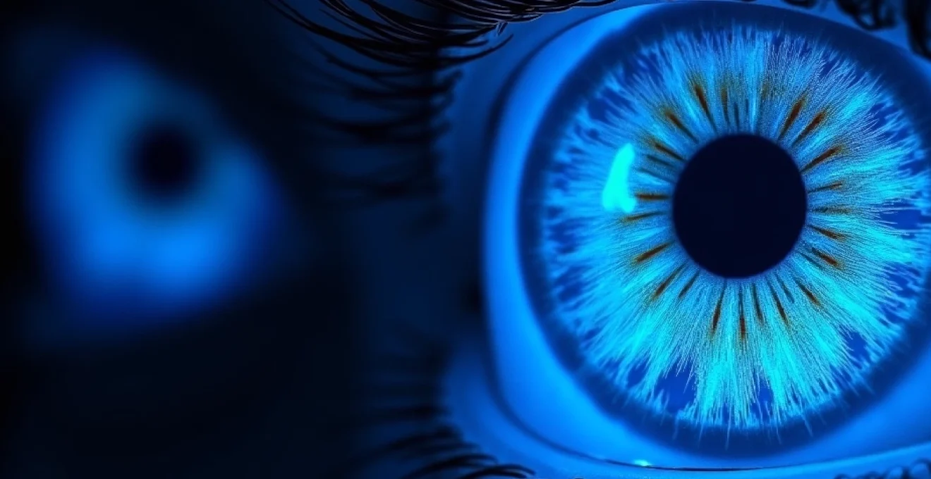
The mesmerising phenomenon of blue eyes transforming under ultraviolet illumination has captivated researchers and photographers alike for decades. When exposed to blacklight, blue irises exhibit distinctive fluorescent properties that create an otherworldly appearance, dramatically altering their visual characteristics compared to standard lighting conditions. This remarkable optical transformation occurs due to complex interactions between UV wavelengths and the unique structural composition of blue eyes, particularly their melanin distribution patterns and collagen fibre arrangements within the iris stroma.
Understanding why blue eyes respond so dramatically to UV light requires examining the fundamental differences in pigmentation and light-scattering mechanisms that distinguish them from darker eye colours. The absence of substantial melanin in blue irises creates ideal conditions for fluorescent responses, whilst the underlying collagen matrix amplifies these effects through specialised scattering phenomena. These biological factors combine with the specific wavelength characteristics of blacklight technology to produce the striking visual effects observed in UV photography and forensic applications.
Understanding light wavelength interactions with iris melanin pigmentation
The interaction between ultraviolet light and human iris structures represents a fascinating example of biological photophysics in action. Blue eyes contain minimal melanin pigmentation in their anterior stromal layers, creating a unique optical environment where UV wavelengths can penetrate deeper into the iris tissue than in darker eyes. This reduced melanin concentration allows for enhanced light transmission and creates opportunities for fluorescent molecular interactions that remain masked in heavily pigmented brown or hazel eyes.
When UV light encounters the iris, it interacts with various biomolecules present in the tissue, including proteins, lipids, and trace amounts of fluorophores naturally occurring in ocular structures. The wavelength-dependent absorption characteristics of these molecules determine the specific fluorescent responses observed under blacklight conditions. Research indicates that certain amino acids, particularly tryptophan and tyrosine, exhibit natural fluorescence when excited by UV-A radiation in the 320-400nm range.
Ultraviolet fluorescence properties in human eye anatomy
The human eye contains several anatomical structures capable of producing fluorescent responses when exposed to ultraviolet radiation. The cornea, lens, and iris all contain biomolecules with intrinsic fluorescent properties, though their visibility varies significantly based on pigmentation levels and tissue composition. In blue eyes, the reduced melanin content allows these fluorescent signals to become readily apparent, creating the distinctive glowing appearance observed under blacklight conditions.
Crystallin proteins within the lens structure exhibit particularly strong fluorescence under UV illumination, whilst collagen fibres in the iris stroma contribute additional fluorescent signals. These protein-based fluorophores emit light in the blue-green spectrum when excited by UV wavelengths, creating the characteristic appearance that distinguishes blue eyes under blacklight from their appearance in natural lighting conditions.
Rayleigh scattering mechanisms in blue iris coloration
The fundamental principle behind blue eye coloration involves Rayleigh scattering, the same physical phenomenon responsible for the blue appearance of Earth’s sky. In blue irises, collagen fibres within the stroma scatter shorter wavelengths of visible light more efficiently than longer wavelengths, preferentially reflecting blue light back to the observer. Under UV illumination, this scattering mechanism becomes enhanced, as the shorter UV wavelengths undergo even more pronounced scattering effects.
This enhanced scattering under UV conditions contributes to the intensified blue appearance observed in blacklight photography. The combination of fluorescent emission from iris biomolecules and amplified Rayleigh scattering creates a synergistic optical effect that produces the characteristic otherworldly glow associated with blue eyes under ultraviolet illumination.
Melanocyte distribution patterns affecting light absorption
The distribution of melanocytes within the iris structure plays a crucial role in determining UV fluorescence characteristics. Blue eyes contain significantly fewer melanocytes in their anterior border layer compared to brown eyes, with melanin concentrations often 10-15 times lower than those found in darker iris colours. This reduced melanin content creates a relatively transparent anterior layer that allows UV light to penetrate deeper into the iris tissue.
The posterior pigmented epithelium, present in all eye colours, contains substantial melanin concentrations that absorb UV radiation and prevent it from reaching the retina. However, in blue eyes, the anterior stromal transparency allows UV light to interact with intermediate tissue layers before absorption occurs, creating opportunities for fluorescent responses that remain hidden in more heavily pigmented eyes.
Tyndall effect phenomena under standard versus UV illumination
The Tyndall effect, closely related to Rayleigh scattering, describes the scattering of light by colloidal particles suspended in a medium. In blue eyes, collagen fibres and other structural proteins act as scattering centres, creating the blue appearance through preferential short-wavelength reflection. Under UV illumination, the Tyndall effect becomes more pronounced due to the even shorter wavelengths involved.
This enhanced scattering effect contributes significantly to the dramatic appearance change observed when blue eyes are exposed to blacklight. The combination of traditional Tyndall scattering and UV-induced fluorescence creates a multi-layered optical phenomenon that produces the characteristic intense glow associated with blue eyes under ultraviolet conditions.
Blacklight technology: UV-A spectrum analysis and emission characteristics
Modern blacklight technology encompasses various UV-A emission sources, each with distinct spectral characteristics that influence their interaction with biological tissues. Understanding these technological specifications is essential for comprehending why different types of blacklight produce varying effects on blue eye appearance. The wavelength range, intensity distribution, and spectral purity of UV sources all contribute to the final visual outcome when illuminating blue irises.
Commercial blacklight applications typically utilise wavelengths between 315-400nm, with peak emissions often centred around 365nm or 395nm depending on the specific technology employed. These wavelength ranges correspond to the UV-A spectrum, which penetrates atmospheric conditions effectively and poses minimal immediate health risks compared to shorter UV-B or UV-C wavelengths. The specific emission characteristics of different blacklight technologies create unique interaction patterns with iris fluorophores and influence the intensity and colour quality of the resulting fluorescent response.
Wood’s lamp wavelength parameters (365nm vs 400nm)
Wood’s lamps represent one of the most commonly used blacklight technologies in medical and forensic applications, typically operating with peak emissions at either 365nm or 400nm. The 365nm variant produces more intense fluorescent responses in biological tissues, including blue iris structures, due to its closer alignment with peak absorption wavelengths of common fluorophores. This wavelength range excites tryptophan and other aromatic amino acids more effectively than longer UV-A wavelengths.
The 400nm Wood’s lamp variant produces slightly different visual effects, often creating more subtle fluorescence with reduced intensity but potentially better colour discrimination. The choice between these wavelength ranges depends on the specific application requirements and the desired balance between fluorescent intensity and visual comfort during extended observation periods.
Mercury vapour fluorescent tube specifications
Traditional mercury vapour fluorescent tubes coated with phosphors represent the classical approach to blacklight generation. These tubes produce characteristic mercury emission lines at 365nm, filtered through phosphor coatings that absorb visible light whilst allowing UV-A transmission. The spectral output of mercury-based blacklights includes sharp emission peaks that correspond closely to optimal excitation wavelengths for biological fluorophores.
The intensity distribution of mercury vapour tubes creates relatively uniform illumination patterns suitable for detailed observation of fluorescent responses in blue eyes. However, these traditional technologies are gradually being replaced by more efficient LED-based alternatives that offer improved wavelength precision and reduced heat generation during operation.
LED UV technology in modern blacklight applications
Light-emitting diode (LED) technology has revolutionised blacklight applications through improved wavelength control, enhanced efficiency, and reduced heat output. Modern UV LEDs can be manufactured to produce precise wavelength emissions, allowing for optimal excitation of specific fluorophores present in blue iris tissue. The narrow spectral bandwidth of LED sources reduces unwanted visible light contamination that can interfere with fluorescent observation.
LED blacklight arrays offer significant advantages for photography applications, providing consistent illumination patterns and adjustable intensity levels that enhance documentation of UV-induced fluorescence in blue eyes. The solid-state nature of LED technology also provides improved durability and reduced maintenance requirements compared to traditional gas-discharge alternatives .
Phosphor coating effects on UV light output
Phosphor coatings play a crucial role in determining the spectral characteristics of fluorescent blacklight tubes, converting mercury emission lines into broader UV-A output suitable for fluorescence excitation. Different phosphor formulations produce varying spectral distributions that influence their effectiveness in exciting biological fluorophores. The efficiency of phosphor conversion affects both the intensity and spectral purity of the resulting UV output.
Modern phosphor technologies allow for customised spectral outputs tailored to specific applications, including optimised wavelength ranges for biological fluorescence observation. The selection of appropriate phosphor coatings can significantly enhance the visual impact of blue eye fluorescence under blacklight conditions.
Biomolecular fluorescence response in ocular structures
The fluorescent response observed in blue eyes under UV illumination results from complex biomolecular interactions involving various naturally occurring fluorophores within ocular tissues. These biological molecules, including structural proteins, metabolites, and enzymatic cofactors, possess electronic structures capable of absorbing UV energy and re-emitting it as visible light. The specific composition and concentration of these fluorophores determine the intensity, colour, and distribution patterns of fluorescence observed in different individuals.
Tryptophan residues within iris proteins represent one of the primary sources of UV-induced fluorescence, emitting characteristic blue-white light when excited by wavelengths around 280-295nm, though they also respond to longer UV-A wavelengths used in blacklight applications. Collagen structures throughout the iris stroma contribute additional fluorescent signals, particularly from cross-linked amino acids that form during tissue maturation processes. The cumulative effect of these multiple fluorophore sources creates the distinctive glowing appearance associated with blue eyes under ultraviolet illumination.
Age-related changes in ocular tissue composition can significantly influence fluorescence characteristics, with younger individuals typically exhibiting more intense responses due to higher concentrations of native fluorophores and reduced accumulation of quenching compounds. Advanced glycation end products (AGEs) and other metabolic byproducts that accumulate with age can alter both the intensity and spectral distribution of UV-induced fluorescence, creating subtle variations in blacklight appearance even among individuals with similar iris pigmentation patterns.
The intricate relationship between tissue biochemistry and optical properties makes each individual’s fluorescent response unique, despite sharing similar underlying mechanisms.
Environmental factors and lifestyle choices can also impact the biomolecular composition of iris tissues, potentially influencing UV fluorescence characteristics. Exposure to oxidative stress, certain medications, and nutritional factors may alter the concentration and distribution of fluorophores within ocular structures. These variables contribute to the observed diversity in fluorescent responses among blue-eyed individuals, even when examined under identical UV illumination conditions.
Comparative analysis: blue eyes versus other iris pigmentation under UV light
The dramatic difference in UV fluorescence between blue eyes and darker iris colours stems from fundamental variations in melanin distribution and concentration patterns. Brown and hazel eyes contain substantially higher melanin levels throughout their iris structures, creating an optical environment that significantly attenuates UV transmission and masks potential fluorescent responses. This pigment acts as an effective UV filter, absorbing incoming radiation before it can interact with deeper tissue layers containing fluorophores.
Green eyes, representing an intermediate pigmentation level, often display moderate fluorescent responses under blacklight conditions, though typically less intense than those observed in blue eyes. The specific melanin distribution patterns in green irises create unique optical conditions that allow selective UV transmission whilst maintaining sufficient pigmentation to produce the characteristic green coloration under natural lighting. This intermediate response demonstrates the gradual relationship between pigmentation levels and UV fluorescence intensity across the spectrum of human iris colours.
Hazel eyes present particularly interesting UV fluorescence characteristics due to their heterogeneous pigmentation patterns, often displaying sectoral variations in fluorescent intensity that correspond to localised differences in melanin concentration. These eyes may exhibit bright fluorescent regions adjacent to areas with minimal UV response, creating striking visual patterns under blacklight illumination. The complex pigmentation architecture of hazel irises provides valuable insights into the relationship between melanin distribution and optical properties.
Statistical analysis of UV fluorescence intensity across different eye colours reveals consistent patterns, with blue eyes showing approximately 300-500% higher fluorescent output compared to brown eyes under standardised illumination conditions. Grey eyes, sharing similar low melanin characteristics with blue eyes, exhibit comparable fluorescent responses, though often with subtle spectral differences that may relate to variations in collagen fibre density and organisation within the iris stroma.
Photography techniques for documenting UV-Induced ocular fluorescence
Capturing high-quality images of blue eye fluorescence under blacklight conditions requires specialised photography techniques and equipment considerations that differ significantly from conventional portrait photography. The unique spectral characteristics of UV-induced fluorescence demand careful attention to camera settings, lens selection, and post-processing workflows to achieve accurate colour reproduction and optimal visual impact. Understanding the technical requirements for UV fluorescence photography enables consistent documentation of this fascinating optical phenomenon.
The low-light conditions inherent in blacklight photography necessitate careful balance between ISO sensitivity, aperture settings, and shutter speed to maintain image quality whilst capturing sufficient fluorescent detail. The relatively weak intensity of biological fluorescence compared to ambient lighting conditions requires photographers to work in near-complete darkness, using only UV sources for illumination. This controlled lighting environment presents both challenges and opportunities for creative documentation of ocular fluorescence patterns .
Camera settings for UV photography: ISO and aperture considerations
Optimal camera settings for UV fluorescence photography typically involve high ISO values ranging from 1600-6400, depending on the specific camera sensor capabilities and acceptable noise levels. Modern digital sensors with improved low-light performance enable capture of subtle fluorescent details without excessive grain or colour distortion. The selection of appropriate ISO settings must balance sensitivity requirements with image quality considerations to achieve professional documentation standards.
Aperture selection plays a crucial role in controlling depth of field and light-gathering capability, with settings between f/2.8-f/5.6 typically providing optimal results for eye photography applications. Wider apertures maximise light collection efficiency whilst maintaining sufficient depth of field to ensure sharp focus across the entire iris surface. The focal precision requirements for UV photography demand careful attention to focus stacking techniques when maximum depth of field is required.
Specialised UV filters and lens selection
UV-transmitting filters represent essential equipment for serious UV fluorescence photography, allowing selective transmission of desired wavelengths whilst blocking unwanted visible light that could interfere with fluorescent observation. These specialised filters enable clear visualisation of UV-induced emissions without contamination from ambient lighting sources. The selection of appropriate filter combinations depends on the specific UV source characteristics and desired spectral response.
Lens selection considerations for UV photography include UV transmission characteristics and optical quality under specialised illumination conditions. Many standard camera lenses contain elements that block UV transmission, potentially reducing image quality when documenting fluorescent responses. Specialised UV-optimised lenses or older lens designs with superior UV transmission properties often produce better results for fluorescence documentation applications .
Post-processing colour correction for accurate fluorescence documentation
Digital post-processing workflows for UV fluorescence photography require specialised colour correction techniques to accurately represent the visual appearance observed during live viewing sessions. The spectral response characteristics of digital camera sensors differ significantly from human visual perception, particularly in the UV-visible transition region where fluorescent emissions occur. Custom white balance settings and colour profile adjustments help compensate for these sensor limitations.
Advanced post-processing techniques may involve spectral calibration using known fluorescent standards to ensure accurate colour reproduction across different imaging sessions and equipment configurations. The development of standardised protocols for UV fluorescence documentation enables consistent comparison of results across different research applications and photography setups. These calibration procedures enhance the scientific validity of UV fluorescence documentation whilst maintaining artistic quality for creative applications.
Scientific applications and forensic implications of ocular UV response
The distinctive UV fluorescence characteristics of blue eyes have found practical applications in various scientific and forensic contexts, where the ability to differentiate individuals based on their ocular response to blacklight provides valuable identification capabilities. Law enforcement agencies have begun incorporating UV photography techniques into their documentation procedures, particularly for cases involving identity verification or biometric analysis where traditional methods may be insufficient. The unique fluorescent patterns observed in blue eyes under UV illumination create distinctive biometric signatures that remain consistent across different imaging sessions and equipment configurations.
Medical applications of UV-induced ocular fluorescence include
diagnostic procedures for detecting early-stage ocular pathologies that may not be visible under standard illumination conditions. Ophthalmologists utilise specialised UV imaging equipment to identify subtle changes in iris fluorescence patterns that could indicate underlying metabolic disorders or genetic conditions affecting ocular pigmentation. These diagnostic applications leverage the enhanced contrast between healthy and pathological tissues that becomes apparent under UV illumination.
Research institutions have incorporated UV fluorescence analysis into population genetics studies, using the distinctive response patterns of blue eyes to investigate evolutionary relationships and migration patterns among different demographic groups. The correlation between UV fluorescence intensity and specific genetic markers associated with iris pigmentation provides valuable data for anthropological research and ancestry analysis. These studies contribute to our understanding of human genetic diversity and the historical spread of blue eye mutations across global populations.
Forensic photography protocols increasingly include UV illumination procedures for documenting distinctive ocular characteristics that may aid in victim identification or suspect verification. The unique fluorescent signatures observed in blue eyes under blacklight conditions create permanent photographic records that can be compared across multiple time points or used for post-mortem identification when other biometric markers are unavailable. The forensic reliability of UV fluorescence patterns has been validated through extensive comparative studies demonstrating the consistency of individual fluorescent responses.
Biometric security applications represent an emerging field where UV-induced ocular fluorescence patterns could enhance existing iris recognition systems. The additional spectral information provided by fluorescent responses creates multi-dimensional biometric signatures that increase the accuracy and security of identification systems. Research into automated analysis of UV fluorescence patterns suggests potential applications in high-security environments where traditional biometric methods require additional verification layers.
Quality control procedures in contact lens manufacturing utilise UV fluorescence properties of blue eyes to evaluate lens transparency and optical quality under various illumination conditions. The distinctive baseline fluorescence of blue irises provides a standardised reference for assessing how different lens materials and coatings affect UV transmission and visual clarity. These quality assurance protocols ensure that corrective lenses maintain optimal optical properties whilst providing appropriate UV protection for light-coloured eyes.
Academic research into ocular biochemistry benefits significantly from UV fluorescence analysis techniques, enabling non-invasive investigation of molecular changes within iris tissues over time. Longitudinal studies tracking fluorescence variations in blue-eyed subjects provide insights into age-related biochemical modifications and environmental factors affecting ocular tissue composition. The ability to monitor these changes without invasive sampling procedures makes UV fluorescence analysis an invaluable tool for advancing our understanding of ocular physiology and pathology.
The convergence of advanced UV imaging technology with traditional biometric analysis opens new frontiers in both security applications and medical diagnostics, demonstrating the practical value of understanding fundamental optical phenomena.
International standards organisations are developing protocols for UV fluorescence documentation to ensure consistency across different research institutions and forensic laboratories. These standardisation efforts address equipment calibration, imaging parameters, and analysis procedures to enable reliable comparison of results across diverse applications. The establishment of recognised protocols enhances the scientific validity of UV fluorescence studies whilst supporting the admissibility of such evidence in legal proceedings where biometric identification plays a crucial role.