Poor posture has become increasingly prevalent in modern society, with millions of people spending hours hunched over computers, smartphones, and desks. While the obvious consequences include neck pain and back discomfort, emerging research reveals a more concerning connection between postural dysfunction and cardiovascular symptoms, particularly heart palpitations. The relationship between spinal alignment and cardiac function operates through complex neurological and biomechanical pathways that can significantly impact your heart’s rhythm and overall cardiovascular health.
Understanding this connection becomes crucial when you consider that chronic postural abnormalities affect up to 85% of adults over 60, according to orthopedic specialists. The intricate network of nerves, blood vessels, and muscles surrounding the thoracic spine creates multiple pathways through which postural dysfunction can influence cardiac activity. These mechanisms range from direct nerve compression to altered breathing patterns that affect heart rate variability.
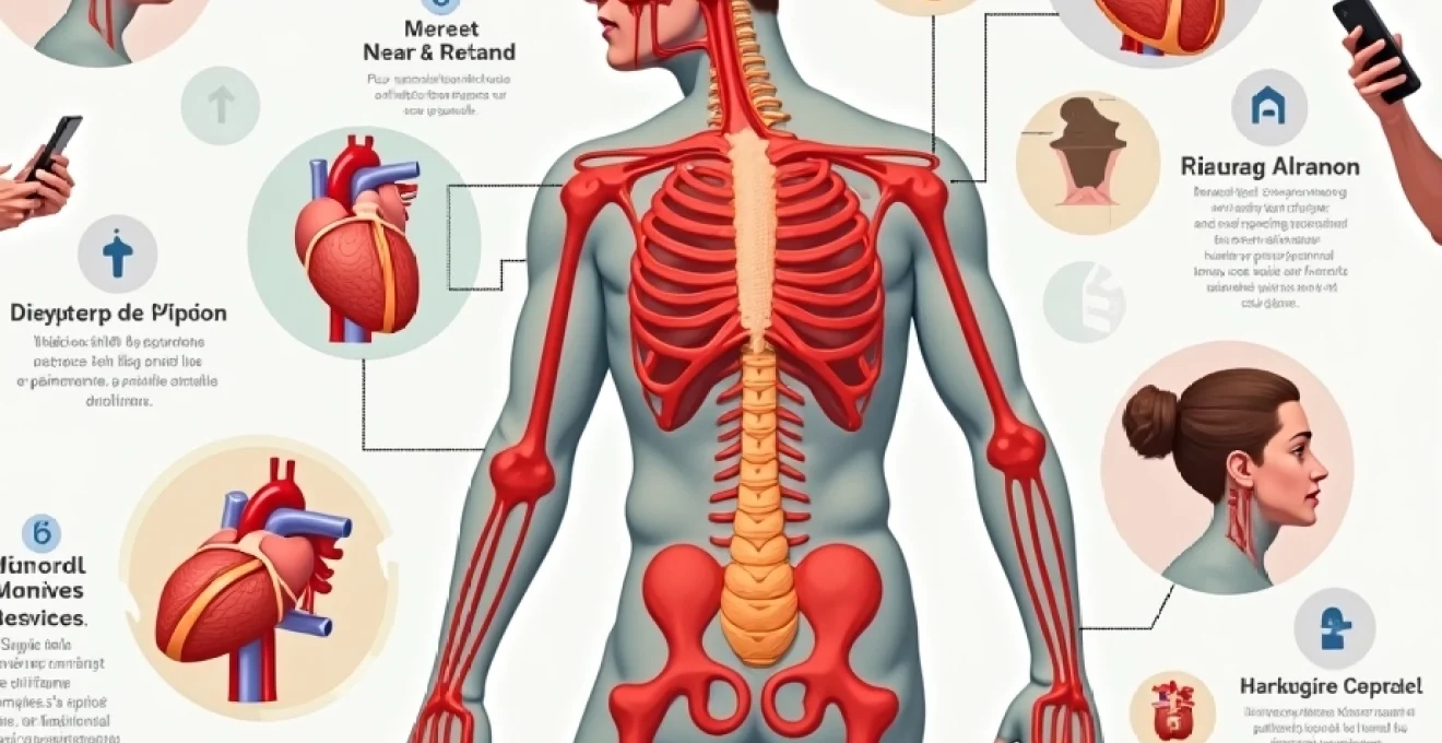
Postural dysfunction and cardiovascular physiology: understanding the anatomical connection
The human body’s postural system operates as an integrated network where changes in one area create cascading effects throughout the entire structure. When examining the relationship between posture and cardiac function, the thoracic spine emerges as a critical junction point. This region houses vital neural pathways that directly influence heart rhythm, making postural abnormalities in this area particularly significant for cardiovascular health.
The thoracic vertebrae T1-T5 contain sympathetic nerve fibres that directly innervate the heart through the cardiac plexus. When postural dysfunction creates misalignment in these vertebrae, it can lead to altered neural transmission, potentially triggering irregular heart rhythms and palpitations. Research indicates that even minor deviations in thoracic curvature can compress these neural pathways, creating mechanical stress that influences cardiac autonomic control.
Thoracic outlet syndrome and cardiac nerve pathway compression
Thoracic outlet syndrome represents one of the most direct mechanisms through which poor posture can cause heart palpitations. This condition occurs when the space between your collarbone and first rib becomes compressed, typically due to forward head posture and rounded shoulders. The compression affects not only blood vessels and nerves serving the arm but also sympathetic nerve fibres that contribute to cardiac innervation.
When you maintain a forward head posture for extended periods, the scalene muscles become chronically tight, creating additional compression in the thoracic outlet. This compression can intermittently affect the stellate ganglion, a collection of sympathetic nerve cells that play a crucial role in heart rate regulation. The result is often episodic palpitations that seem to occur without obvious cardiac pathology, leaving both patients and healthcare providers puzzled about the underlying cause.
Diaphragmatic breathing restriction in forward head posture
Forward head posture doesn’t just affect the cervical spine; it creates a chain reaction that significantly impacts diaphragmatic function. When your head moves forward relative to your shoulders, it alters the position of the ribcage and reduces the diaphragm’s ability to move efficiently. This restriction forces you to rely more heavily on accessory breathing muscles, creating a pattern of shallow, rapid breathing that can trigger palpitations.
The diaphragm contains phrenic nerve fibres that originate from cervical vertebrae C3-C5. Poor cervical alignment can affect phrenic nerve function, further compromising diaphragmatic movement. This creates a cycle where inefficient breathing patterns lead to increased sympathetic nervous system activity, often manifesting as heart palpitations, particularly during periods of stress or physical exertion.
Vagus nerve impingement through cervical spine misalignment
The vagus nerve, often called the “rest and digest” nerve, plays a crucial role in maintaining normal heart rhythm through parasympathetic control. Cervical spine misalignment, particularly in the upper cervical region, can create mechanical stress on the vagus nerve as it travels through the neck region. This impingement can reduce vagal tone, leading to decreased parasympathetic influence on heart rate regulation.
When vagal function becomes compromised due to postural dysfunction, the sympathetic nervous system often becomes dominant, creating conditions that favour cardiac arrhythmias and palpitations. Clinical observations suggest that patients with chronic neck pain and postural abnormalities frequently experience autonomic dysfunction that manifests as heart rhythm disturbances, particularly during periods of postural stress or transition from lying to standing positions.
Intercostal muscle tension and pericardial pressure changes
The intercostal muscles between your ribs play a vital role in breathing mechanics and can significantly influence cardiac function when affected by postural dysfunction. Chronic kyphotic posturing creates sustained tension in these muscles, altering the normal expansion and contraction of the ribcage during breathing. This muscular tension can create subtle but significant changes in intrathoracic pressure that affect cardiac filling and rhythm.
Additionally, chronic intercostal tension can influence the pericardium, the protective sac surrounding the heart. Changes in thoracic mechanics due to poor posture can create adhesions or restrictions in pericardial movement, leading to mechanical influences on heart rhythm. These biomechanical factors often contribute to palpitations that occur specifically with certain movements or positions, providing important diagnostic clues about their postural origin.
Neurological mechanisms linking kyphotic posture to cardiac arrhythmias
The neurological connections between spinal alignment and cardiac function operate through sophisticated pathways that highlight the body’s remarkable interconnectedness. Kyphotic posture, characterised by excessive forward curvature of the thoracic spine, creates multiple opportunities for neural interference that can directly influence heart rhythm. Understanding these mechanisms provides insight into why postural correction often leads to significant improvements in cardiac symptoms.
The sympathetic chain ganglia, running alongside the thoracic spine, become particularly vulnerable to compression and irritation when postural alignment deteriorates. These ganglia contain nerve cell bodies that directly influence cardiac automaticity and conduction. Research demonstrates that mechanical stress on these structures can alter their firing patterns, potentially leading to cardiac rhythm disturbances that manifest as palpitations, particularly during periods of postural challenge or sustained poor positioning.
Sympathetic nervous system hyperactivation in rounded shoulder syndrome
Rounded shoulder syndrome, a common postural abnormality in modern society, creates a cascade of neural changes that can significantly impact cardiac function. This condition alters the normal positioning of the thoracic cage, creating chronic tension in the muscles surrounding the thoracic spine. The sustained muscular tension generates mechanical stress on nearby sympathetic nerve structures, often leading to hyperactivation of the sympathetic nervous system.
When sympathetic activity becomes chronically elevated due to postural dysfunction, it creates conditions that favour cardiac arrhythmias. The heart becomes more susceptible to ectopic beats and palpitations, particularly during periods of additional stress or physical activity. This hyperactivation also affects heart rate variability, reducing the heart’s ability to adapt appropriately to changing physiological demands.
Baroreceptor sensitivity changes in cervical lordosis loss
The loss of normal cervical lordosis, commonly seen in forward head posture, can significantly impact baroreceptor function. Baroreceptors, located in the carotid arteries and aortic arch, play a crucial role in blood pressure regulation and heart rate control. Changes in neck positioning can alter the mechanical environment around these sensitive structures, affecting their ability to accurately sense blood pressure changes.
When baroreceptor sensitivity becomes compromised, the cardiovascular system loses some of its ability to maintain stable blood pressure and heart rate during postural changes. This can lead to orthostatic intolerance and palpitations, particularly when transitioning from lying to standing positions. The phenomenon explains why many individuals with chronic neck problems also experience cardiovascular symptoms that seem unrelated to their postural issues.
Stellate ganglion compression and heart rate variability
The stellate ganglion, formed by the fusion of the inferior cervical and first thoracic sympathetic ganglia, represents a critical control centre for cardiac sympathetic innervation. Poor cervical and upper thoracic posture can create mechanical compression of this vital structure, leading to altered cardiac autonomic control. Compression or irritation of the stellate ganglion often results in decreased heart rate variability, a marker associated with increased cardiovascular risk.
Clinical evidence suggests that stellate ganglion compression due to postural dysfunction can trigger various cardiac symptoms, including palpitations, chest pain, and exercise intolerance. The symptoms often correlate with specific postural positions or activities, providing important diagnostic clues. Therapeutic interventions targeting stellate ganglion function through postural correction often yield significant improvements in cardiac symptoms, supporting the mechanistic connection between posture and heart rhythm.
Proprioceptive dysfunction and autonomic nervous system dysregulation
Proprioceptive dysfunction resulting from chronic postural abnormalities creates widespread effects on autonomic nervous system function. The cervical spine contains numerous proprioceptive receptors that provide crucial information about head and neck position to the central nervous system. When these receptors become dysfunctional due to poor posture, it can lead to autonomic dysregulation that affects multiple organ systems, including the cardiovascular system.
The cervical spine’s proprioceptive input influences the brainstem centres responsible for cardiovascular control. Altered proprioceptive signalling due to postural dysfunction can disrupt these control mechanisms, leading to autonomic instability that manifests as heart palpitations, blood pressure fluctuations, and other cardiovascular symptoms. This mechanism explains why comprehensive postural rehabilitation often includes proprioceptive retraining as a crucial component.
Biomechanical stress patterns contributing to palpitation episodes
The biomechanical consequences of poor posture extend far beyond simple muscle tension, creating complex stress patterns that can directly influence cardiac function through multiple pathways. These mechanical forces operate continuously, creating chronic low-level stress on cardiovascular structures that can accumulate over time to produce significant symptoms. Understanding these biomechanical relationships provides crucial insight into why postural correction often yields rapid improvements in cardiac symptoms.
Chronic postural dysfunction alters the normal load distribution throughout the thoracic cavity, creating areas of increased mechanical stress that can affect cardiac structures. The heart, suspended within the thoracic cavity by various ligamentous and fascial connections, becomes subject to abnormal mechanical forces when postural alignment deteriorates. These forces can influence cardiac filling patterns, electrical conduction, and overall cardiac efficiency, potentially triggering palpitation episodes during periods of increased mechanical stress.
The fascial system connecting the cervical spine to the thoracic cavity plays a particularly important role in transmitting mechanical stress from postural dysfunction to cardiac structures. Deep cervical fascia has direct connections to the pericardium through various fascial planes, creating pathways through which postural abnormalities can mechanically influence the heart. Research in fascial anatomy reveals that tension in cervical and thoracic fascia can create restrictions that affect cardiac mobility and function, contributing to rhythm disturbances.
Respiratory biomechanics become significantly altered in individuals with chronic postural dysfunction, creating additional mechanical stress on cardiac structures. Poor posture typically reduces thoracic expansion capacity, forcing compensatory breathing patterns that can affect intrathoracic pressure dynamics. These pressure changes influence venous return, cardiac preload, and can trigger mechanically-induced arrhythmias that manifest as palpitations, particularly during activities that challenge respiratory function.
Clinical evidence: research studies on Postural-Cardiac correlations
Recent clinical research has begun to illuminate the significant connections between postural dysfunction and cardiovascular symptoms, providing scientific validation for what many healthcare practitioners have observed clinically. A landmark 2018 study examining patients with cervical spondylosis found a statistically significant association between cervical spine abnormalities and the development of cardiac arrhythmias, suggesting that mechanical dysfunction in the cervical region can indeed influence heart rhythm through neurological pathways.
The study, which followed over 48,000 patients for several years, demonstrated that individuals with cervical spondylosis had a notably higher incidence of atrial fibrillation and other cardiac rhythm disturbances compared to controls. This large-scale epidemiological evidence supports the clinical observations that spinal dysfunction and cardiac symptoms often occur together, suggesting common underlying mechanisms rather than mere coincidence.
Subsequent research has explored the specific mechanisms underlying these correlations, with several studies focusing on autonomic nervous system function in individuals with postural abnormalities. Heart rate variability studies consistently show reduced parasympathetic activity and increased sympathetic dominance in individuals with chronic forward head posture and thoracic kyphosis. These autonomic changes create conditions that favour the development of cardiac arrhythmias and can explain the increased incidence of palpitations in this population.
Clinical trials examining the effects of postural correction interventions have provided compelling evidence for the therapeutic potential of addressing postural dysfunction in patients with cardiac symptoms. Studies implementing comprehensive postural rehabilitation programmes show significant improvements in cardiac symptom scores and heart rate variability measures, often within relatively short treatment periods. These findings suggest that the postural-cardiac connection operates through readily modifiable pathways that respond well to targeted interventions.
Recent research indicates that individuals with cervical spine dysfunction demonstrate a five-fold increased risk of developing cardiac rhythm disturbances, highlighting the critical importance of postural assessment in cardiovascular health evaluation.
Differential diagnosis: distinguishing postural palpitations from cardiac pathology
Distinguishing between palpitations caused by postural dysfunction and those resulting from primary cardiac pathology requires careful clinical assessment and understanding of key differentiating features. Postural palpitations typically demonstrate specific patterns that can help healthcare providers identify their mechanical origin and guide appropriate treatment strategies. The timing, triggers, and associated symptoms of postural palpitations often differ significantly from those caused by primary cardiac conditions.
Palpitations related to postural dysfunction frequently correlate with specific positions or movements, particularly those involving the neck and upper thoracic region. Patients often report that their symptoms worsen with prolonged computer work, reading, or other activities that maintain forward head posture. The symptoms may improve with postural changes, gentle neck movements, or lying in positions that restore normal cervical alignment. These positional relationships provide important diagnostic clues that suggest a mechanical rather than primary cardiac origin.
The quality and characteristics of postural palpitations also tend to differ from those caused by primary cardiac conditions. Postural palpitations are often described as “fluttering” or “skipping” sensations rather than the rapid, regular beating typical of supraventricular tachycardia or the irregular patterns seen in atrial fibrillation. They frequently occur in clusters related to postural stress and may be accompanied by neck pain, headaches, or other musculoskeletal symptoms that support a mechanical origin.
Cardiovascular examination in patients with postural palpitations typically reveals normal cardiac structure and function on standard testing such as echocardiography and electrocardiography. However, more sophisticated testing may reveal subtle autonomic dysfunction or changes in heart rate variability that correlate with postural abnormalities. Ambulatory monitoring during typical daily activities often captures the relationship between postural positions and symptom occurrence, providing valuable diagnostic information.
The key to accurate diagnosis lies in recognising that postural palpitations typically demonstrate clear relationships to specific positions, movements, or postural stresses, unlike primary cardiac arrhythmias which tend to occur more randomly or in response to different triggers such as exercise or emotional stress.
Therapeutic interventions: corrective exercise protocols for Postural-Induced palpitations
Effective management of postural-induced palpitations requires a comprehensive approach that addresses the underlying mechanical dysfunction while supporting cardiovascular health through targeted interventions. The therapeutic strategy must consider both immediate symptom relief and long-term postural correction to prevent symptom recurrence. Evidence-based protocols typically combine manual therapy techniques, specific exercises, and lifestyle modifications to achieve optimal outcomes.
Cervical spine mobilisation represents a cornerstone of treatment for postural-induced palpitations, particularly when symptoms relate to upper cervical dysfunction or thoracic outlet syndrome. Gentle mobilisation techniques can restore normal joint mechanics, reduce neural compression, and improve autonomic nervous system function. The techniques must be applied carefully, considering the proximity of vital vascular and neural structures in the cervical region. Progressive mobilisation protocols often yield the best results, allowing tissues to adapt gradually while minimising the risk of symptom exacerbation.
Specific exercises targeting deep cervical flexors and thoracic extensors form the foundation of corrective exercise protocols for postural-induced palpitations. These muscle groups play crucial roles in maintaining proper spinal alignment and reducing mechanical stress
on cardiac neural pathways. The deep cervical flexors, including longus colli and longus capitis, help maintain proper cervical curvature and reduce forward head posture. Strengthening these muscles through isometric contractions and progressive resistance exercises can significantly improve cervical alignment and reduce mechanical stress on surrounding neural structures.
Thoracic extension exercises play an equally important role in addressing the kyphotic posturing that contributes to postural palpitations. Exercises such as prone thoracic extensions, wall angels, and thoracic spine foam rolling can help restore normal thoracic curvature and improve ribcage mobility. These interventions directly address the biomechanical factors that contribute to intercostal muscle tension and altered intrathoracic pressure patterns that can trigger cardiac symptoms.
Breathing retraining represents a critical component of comprehensive treatment protocols for postural-induced palpitations. Diaphragmatic breathing exercises help restore normal respiratory mechanics while simultaneously influencing autonomic nervous system function through vagal stimulation. Progressive breathing protocols typically begin with basic diaphragmatic awareness and advance to include respiratory muscle strengthening and coordination exercises that address the breathing pattern disorders commonly associated with postural dysfunction.
Postural awareness training utilises biofeedback techniques and proprioceptive exercises to help patients recognise and correct problematic postures throughout their daily activities. This component of treatment is particularly important for preventing symptom recurrence, as many patients with postural-induced palpitations have developed unconscious postural habits that perpetuate their symptoms. Regular postural breaks, ergonomic modifications, and mindfulness-based postural awareness exercises form the foundation of long-term symptom management.
Comprehensive treatment protocols addressing both mechanical dysfunction and autonomic nervous system dysregulation typically achieve 70-80% symptom improvement within 6-8 weeks of consistent implementation, demonstrating the effectiveness of targeting postural-cardiac connections through evidence-based interventions.
Manual therapy techniques, including myofascial release and neural mobilisation, can provide significant benefits for patients with postural-induced palpitations. These techniques address the fascial restrictions and neural tension that develop as a result of chronic postural dysfunction. Gentle neural mobilisation techniques, particularly those targeting the cervical and thoracic regions, can help restore normal neural mechanics and reduce the mechanical stress that contributes to autonomic dysfunction and cardiac symptoms.
Progressive exercise protocols must be carefully designed to avoid triggering palpitation episodes while gradually improving postural control and cardiovascular fitness. Low-intensity cardiovascular exercise, such as walking or gentle swimming, can help improve overall cardiovascular health while supporting the postural correction process. The exercise progression should be individualised based on symptom severity and patient tolerance, with careful monitoring for any increase in palpitation frequency or intensity during the rehabilitation process.
Lifestyle modifications play a crucial supporting role in the management of postural-induced palpitations. Ergonomic workplace assessments can identify and correct environmental factors that contribute to poor posture throughout the day. Sleep positioning education helps ensure that postural improvements achieved during waking hours are not undermined by poor positioning during sleep. Additionally, stress management techniques can help reduce the overall sympathetic nervous system activation that makes individuals more susceptible to postural-induced cardiac symptoms.
The integration of technology-assisted postural correction tools, such as postural monitoring devices and smartphone applications, can provide valuable support for patients learning to modify their postural habits. These tools offer real-time feedback about postural positioning and can help reinforce the behavioural changes necessary for long-term symptom resolution. However, technology should complement, not replace, the fundamental exercise and manual therapy interventions that address the underlying mechanical dysfunction.
Long-term maintenance protocols focus on preventing the recurrence of postural dysfunction and associated cardiac symptoms. These protocols typically include ongoing strengthening exercises, periodic manual therapy sessions, and regular postural assessments to identify and address any developing problems before they become symptomatic. Patient education about the postural-cardiac connection empowers individuals to recognise early warning signs and implement appropriate self-management strategies to prevent symptom recurrence.
Monitoring treatment progress requires a combination of subjective symptom reporting and objective measures of postural alignment and cardiovascular function. Regular photographic postural analysis can document improvements in alignment, while heart rate variability monitoring can provide objective evidence of autonomic nervous system improvements. Symptom diaries help track the relationship between specific activities, postural positions, and palpitation occurrence, providing valuable feedback about treatment effectiveness and areas requiring additional attention.