Post-cholecystectomy diarrhoea affects approximately 10-20% of patients following gallbladder removal surgery, presenting a significant challenge for both patients and healthcare providers. This troublesome complication can persist for weeks or even months after what is typically considered a routine surgical procedure. The condition occurs due to fundamental changes in bile acid metabolism and intestinal function following the removal of the gallbladder’s storage capacity. Understanding the underlying mechanisms and implementing appropriate treatment strategies is crucial for restoring normal bowel function and improving quality of life for affected patients.
Understanding Post-Cholecystectomy diarrhoea: bile acid malabsorption mechanisms
The development of chronic diarrhoea following gallbladder removal stems from complex alterations in bile acid physiology. Without the gallbladder’s storage and concentration functions, bile acids flow continuously into the small intestine rather than being released in response to meals. This continuous flow disrupts the normal enterohepatic circulation, leading to bile acid malabsorption and subsequent colonic irritation.
Bile acid dysregulation following laparoscopic cholecystectomy
Laparoscopic cholecystectomy, whilst minimally invasive, fundamentally alters bile acid homeostasis. The absence of gallbladder contractions means bile acids enter the duodenum at suboptimal concentrations and inappropriate timing. Primary bile acids such as cholic acid and chenodeoxycholic acid become dysregulated, affecting their normal absorption in the terminal ileum. This dysregulation creates a cascade effect, influencing both digestive efficiency and bowel motility patterns.
Primary bile acid diarrhoea vs secondary BAM syndrome
Post-cholecystectomy diarrhoea primarily manifests as secondary bile acid malabsorption (BAM) syndrome, distinct from primary bile acid diarrhoea. Secondary BAM results from structural or functional changes to the ileum’s bile acid transport mechanisms following surgery. The condition differs from primary BAM, which involves inherited defects in bile acid synthesis. Understanding this distinction is crucial for selecting appropriate therapeutic interventions and predicting treatment outcomes.
Hepatic bile production changes after gallbladder removal
Following cholecystectomy, the liver attempts to compensate for the loss of bile storage capacity by increasing bile acid synthesis. This compensatory mechanism often proves inadequate, leading to bile acid deficiency during peak digestive periods and excess during fasting states. The hepatic response involves upregulation of 7α-hydroxylase, the rate-limiting enzyme in bile acid synthesis, but this adaptation may take several months to establish optimal balance.
Colonic mucosa response to increased bile salt concentrations
Elevated bile salt concentrations in the colon trigger inflammatory responses and alter water and electrolyte transport mechanisms. The colonic mucosa becomes increasingly permeable, leading to secretory diarrhoea characterised by high-volume, watery stools. Secondary bile acids, particularly deoxycholic acid produced by bacterial 7α-dehydroxylation, exert potent secretory effects on colonocytes. This process explains why post-cholecystectomy diarrhoea often presents with urgency and frequent, voluminous bowel movements.
Pharmacological interventions for Post-Cholecystectomy diarrhoea management
Effective pharmacological management requires a comprehensive approach targeting bile acid sequestration, bowel motility regulation, and microbiome restoration. The selection of appropriate medications depends on symptom severity, patient tolerance, and underlying comorbidities. Treatment protocols should be individualised and regularly monitored for efficacy and adverse effects.
Bile acid sequestrants: cholestyramine and colesevelam protocols
Bile acid sequestrants represent the first-line pharmacological intervention for post-cholecystectomy diarrhoea. Cholestyramine, administered at doses of 4-8 grams twice daily before meals, binds excess bile acids in the intestinal lumen, preventing colonic irritation. Colesevelam offers improved tolerability with fewer gastrointestinal side effects and can be administered as 625mg tablets, typically three to six tablets daily. Both medications require careful timing to avoid interference with the absorption of other medications, particularly fat-soluble vitamins and cardiac medications.
The efficacy of bile acid sequestrants in managing post-cholecystectomy diarrhoea ranges from 70-85%, with symptom improvement typically observed within 2-4 weeks of initiating treatment.
Loperamide hydrochloride dosage optimisation for BAD
Loperamide hydrochloride provides symptomatic relief by reducing intestinal motility and enhancing water absorption in the colon. For post-cholecystectomy diarrhoea, higher doses than those used for acute diarrhoea may be necessary, with some patients requiring 8-16mg daily divided into multiple doses. The medication’s opioid receptor agonist properties help normalise transit time and stool consistency. Careful monitoring is essential to prevent constipation or toxic megacolon, particularly in elderly patients or those with underlying intestinal pathology.
Rifaximin treatment for small intestinal bacterial overgrowth
Small intestinal bacterial overgrowth (SIBO) commonly develops following cholecystectomy due to altered bile flow and reduced antimicrobial activity. Rifaximin, a non-absorbable antibiotic, effectively targets pathogenic bacteria whilst preserving beneficial colonic microflora. Treatment protocols typically involve 400mg three times daily for 10-14 days, with some patients requiring repeated courses. The medication’s local action minimises systemic side effects whilst addressing the bacterial dysbiosis contributing to persistent diarrhoeal symptoms.
Probiotics supplementation: lactobacillus plantarum and bifidobacterium strains
Targeted probiotic supplementation can help restore intestinal microbiome balance disrupted by altered bile acid metabolism. Lactobacillus plantarum demonstrates particular efficacy in bile acid deconjugation and inflammatory modulation. Bifidobacterium strains, including B. longum and B. bifidum, support colonocyte health and enhance short-chain fatty acid production. Multi-strain formulations containing 10-50 billion colony-forming units daily provide optimal therapeutic benefits when administered consistently for 8-12 weeks.
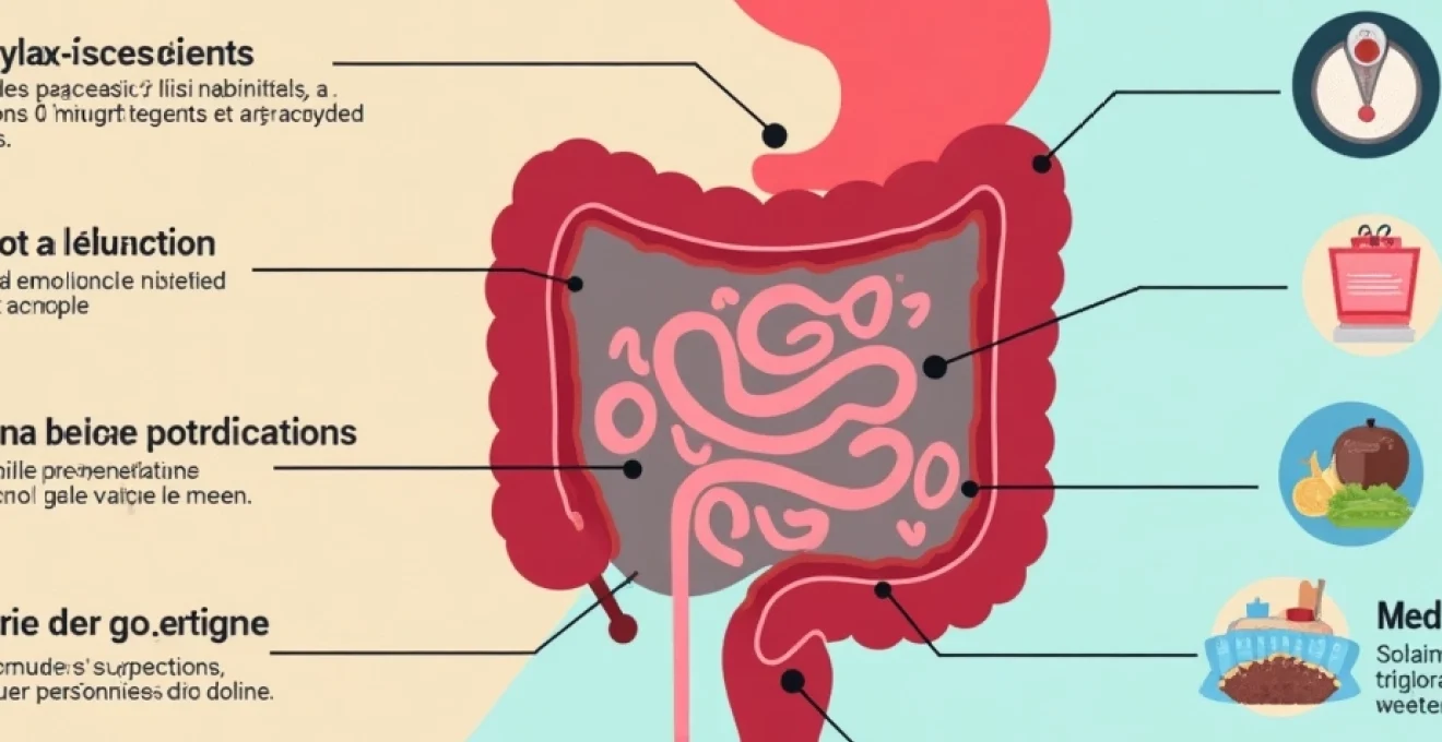
Dietary modifications and nutritional strategies for bile acid diarrhoea
Comprehensive dietary management forms the cornerstone of post-cholecystectomy diarrhoea treatment, often providing significant symptom relief when implemented correctly. The approach focuses on reducing bile acid stimulation whilst maintaining adequate nutrition and preventing malabsorption of essential nutrients. Patients require careful guidance to navigate dietary restrictions without compromising nutritional status.
Low-fat diet implementation: reducing triglyceride intake below 40g daily
Implementing a low-fat diet effectively reduces bile acid demand and subsequent colonic irritation. Daily fat intake should be limited to 30-40 grams, distributed across multiple small meals to optimise digestion. This restriction requires careful meal planning and label reading, as hidden fats in processed foods can significantly contribute to total daily intake. Patients should focus on lean proteins, complex carbohydrates, and fat-free or low-fat dairy products whilst avoiding fried foods, fatty meats, and high-fat desserts.
Soluble fibre supplementation: psyllium husk and methylcellulose
Soluble fibre supplementation helps bind excess bile acids and improve stool consistency without exacerbating diarrhoeal symptoms. Psyllium husk, administered as 5-10 grams daily in divided doses, provides excellent bile acid sequestration whilst promoting beneficial bacterial growth. Methylcellulose offers an alternative for patients intolerant to psyllium, with similar efficacy in stool normalisation. Gradual introduction and adequate fluid intake prevent gastrointestinal discomfort and ensure optimal therapeutic benefits.
Medium-chain triglycerides (MCT) oil integration
Medium-chain triglycerides bypass normal fat digestion pathways, providing essential fatty acids without stimulating excessive bile acid release. MCT oil can be incorporated into the diet starting with 1-2 teaspoons daily and gradually increasing to 1-2 tablespoons as tolerated. This approach maintains caloric intake whilst reducing symptoms associated with long-chain fatty acid consumption. MCT oil’s rapid absorption and hepatic metabolism make it particularly beneficial for patients with severe fat malabsorption.
Calcium and magnesium balance for stool consistency
Maintaining appropriate calcium and magnesium balance significantly impacts stool consistency and bowel movement frequency. Calcium supplementation at 500-1000mg daily can help bind fatty acids and reduce stool looseness, particularly when combined with bile acid sequestrants. Magnesium levels require careful monitoring, as deficiency can worsen diarrhoea whilst excess may have laxative effects. Regular blood tests and symptom assessment guide optimal dosing strategies for individual patients.
Patients following comprehensive dietary modification protocols report symptom improvement in 65-80% of cases within 4-6 weeks of implementation.
Advanced diagnostic approaches for Post-Operative diarrhoea assessment
Accurate diagnosis of post-cholecystectomy diarrhoea requires comprehensive evaluation to exclude other gastrointestinal pathologies and confirm bile acid malabsorption. Advanced diagnostic techniques help differentiate between various causes of chronic diarrhoea and guide targeted treatment approaches. The diagnostic workup should be systematic and evidence-based to avoid unnecessary investigations whilst ensuring accurate diagnosis.
SeHCAT (selenium homocholic acid taurine) scanning represents the gold standard for diagnosing bile acid malabsorption, measuring bile acid retention at seven days post-administration. Normal retention exceeds 15%, with values below 10% indicating severe malabsorption requiring aggressive treatment. Faecal bile acid quantification provides an alternative diagnostic approach when SeHCAT scanning is unavailable, though it requires multiple stool collections and may be less convenient for patients.
Comprehensive stool analysis should include microscopy, culture, and inflammatory markers to exclude infectious or inflammatory causes of persistent diarrhoea. Faecal calprotectin levels help differentiate between functional and organic causes, with elevated levels suggesting inflammatory bowel disease or microscopic colitis. Additional testing may include coeliac serology, thyroid function, and vitamin B12 levels to identify concurrent conditions contributing to gastrointestinal symptoms.
Hydrogen breath testing can identify small intestinal bacterial overgrowth, which frequently complicates post-cholecystectomy syndrome. Lactulose or glucose breath tests demonstrate bacterial fermentation patterns and guide antibiotic treatment decisions. Colonoscopy with biopsies may be indicated for patients with alarm symptoms or those failing to respond to standard treatments, particularly to exclude microscopic colitis or other structural abnormalities.
Long-term prognosis and specialist referral pathways
The long-term prognosis for post-cholecystectomy diarrhoea varies significantly among patients, with approximately 60-70% experiencing substantial improvement within six months of implementing appropriate treatment strategies. Factors influencing outcomes include symptom severity at presentation, patient compliance with treatment protocols, and the presence of concurrent gastrointestinal conditions. Early intervention and comprehensive management approaches generally yield superior results compared to delayed treatment initiation.
Most patients achieve satisfactory symptom control through combination therapy involving dietary modifications, bile acid sequestrants, and supportive medications. However, approximately 15-20% of patients experience persistent or refractory symptoms requiring specialist gastroenterology input. These patients may benefit from advanced therapeutic options including bile acid receptor agonists, specialized dietary interventions, or investigational treatments currently under development.
Specialist referral should be considered for patients with severe, persistent symptoms lasting more than three months despite appropriate initial treatment. Additional indications include significant weight loss, nutritional deficiencies, or quality of life impairment interfering with daily activities. Gastroenterologists can provide access to advanced diagnostic techniques, specialized medications, and clinical trials investigating novel therapeutic approaches for refractory post-cholecystectomy syndrome.
Studies indicate that 85-90% of patients with post-cholecystectomy diarrhoea achieve acceptable symptom control within 12 months when managed through structured, multidisciplinary care pathways.
Regular follow-up appointments should be scheduled to monitor treatment response, adjust medications, and screen for complications such as nutritional deficiencies or medication side effects. Vitamin D, B12, and fat-soluble vitamin levels require periodic monitoring, particularly in patients using bile acid sequestrants long-term. Patient education regarding symptom recognition and treatment adherence remains crucial for maintaining optimal outcomes and preventing symptom recurrence.
Emergency interventions for severe Post-Cholecystectomy diarrhoea complications
Severe post-cholecystectomy diarrhoea can occasionally lead to life-threatening complications requiring immediate medical intervention. Dehydration and electrolyte imbalances represent the most common emergency presentations, particularly in elderly patients or those with limited physiological reserves. Recognition of warning signs and prompt treatment initiation are essential for preventing progression to more serious complications such as acute kidney injury or cardiac arrhythmias.
Fluid resuscitation protocols should focus on rapid restoration of intravascular volume whilst addressing electrolyte abnormalities, particularly sodium, potassium, and magnesium depletion. Intravenous fluid therapy with balanced crystalloid solutions provides optimal electrolyte replacement, avoiding the complications associated with normal saline administration. Severe cases may require central venous access and intensive monitoring to guide fluid management and prevent volume overload.
Emergency pharmacological interventions may include high-dose loperamide administration, temporary bile acid sequestrant therapy, and antimicrobial treatment for suspected bacterial overgrowth. Octreotide, a synthetic somatostatin analogue, can provide rapid symptom relief in severe cases by reducing intestinal secretions and motility. However, this medication requires careful dosing and monitoring due to potential side effects including gallstone formation and glucose intolerance.
Nutritional support becomes critical during severe exacerbations, with some patients requiring parenteral nutrition to maintain caloric intake and prevent further weight loss. Enteral nutrition through nasogastric or percutaneous endoscopic gastrostomy tubes may be considered for patients unable to maintain oral intake due to persistent nausea or vomiting. Close collaboration between gastroenterology, nutrition, and critical care teams ensures comprehensive management of complex cases requiring intensive interventions.