Experiencing regurgitation or “throwing up in your mouth” can be an unsettling and uncomfortable sensation that affects millions of people worldwide. This phenomenon, medically known as acid reflux or gastroesophageal reflux, involves the involuntary return of stomach contents into the oesophagus and sometimes into the mouth. Unlike full vomiting episodes, this condition often presents as a sudden backflow of acidic material that creates a burning sensation and unpleasant taste. Understanding the underlying mechanisms behind this digestive disruption is crucial for identifying appropriate treatment strategies and preventing potential complications. The causes range from temporary lifestyle factors to serious underlying medical conditions that require professional intervention.
Gastroesophageal reflux disease (GORD) and acid regurgitation mechanisms
Gastroesophageal reflux disease represents the most common cause of acid regurgitation, affecting approximately 20% of the adult population in developed countries. This chronic condition occurs when stomach acid regularly flows back into the oesophagus, creating symptoms that extend beyond occasional heartburn. The pathophysiology involves complex interactions between gastric pressure, oesophageal motility, and sphincter function that normally prevent retrograde movement of gastric contents.
The severity of GORD symptoms correlates directly with the frequency and duration of acid exposure to oesophageal tissues. Research indicates that patients experiencing daily regurgitation episodes face significantly higher risks of developing erosive oesophagitis and other serious complications. The acidic nature of regurgitated material, typically containing hydrochloric acid with a pH below 2.0, creates immediate tissue damage upon contact with the sensitive oesophageal lining.
Lower oesophageal sphincter dysfunction and transient relaxations
The lower oesophageal sphincter (LES) functions as the primary barrier preventing gastric reflux, maintaining baseline pressures between 10-30 mmHg in healthy individuals. Dysfunction occurs through two primary mechanisms: permanently reduced sphincter pressure or inappropriate transient lower oesophageal sphincter relaxations (TLESRs). These transient relaxations account for approximately 95% of reflux episodes in patients with normal LES pressure, lasting 10-60 seconds and allowing rapid equalisation of gastric and oesophageal pressures.
Factors contributing to LES dysfunction include dietary triggers such as chocolate, caffeine, and fatty foods, which directly reduce sphincter pressure through hormonal mechanisms. Additionally, medications including calcium channel blockers, nitrates, and anticholinergics can significantly impair LES function, necessitating careful medication review in patients experiencing frequent regurgitation episodes.
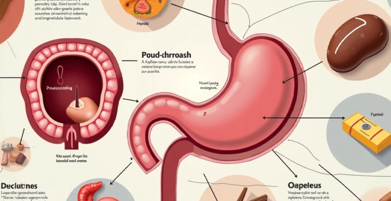
Hiatal hernia impact on gastric content reflux
Hiatal hernias, present in approximately 15% of adults over 50 years, substantially increase the likelihood of acid regurgitation by disrupting normal anatomical relationships. The herniation of gastric tissue through the diaphragmatic hiatus compromises the natural anti-reflux mechanisms, including the acute angle of His and the diaphragmatic crura support. Large hiatal hernias create a gastric reservoir above the diaphragm, facilitating gravitational reflux regardless of LES competency.
The relationship between hiatal hernia size and symptom severity demonstrates significant individual variation, with some patients experiencing severe regurgitation despite small hernias, whilst others remain asymptomatic with substantial anatomical distortion. Sliding hiatal hernias represent the most common type, accounting for 95% of cases and directly correlating with increased acid exposure times during oesophageal pH monitoring studies.
Pepsin and hydrochloric acid composition in regurgitated material
The biochemical composition of regurgitated material provides important diagnostic information and influences symptom severity. Gastric juice contains pepsinogen, which converts to active pepsin in acidic environments, creating a proteolytic enzyme capable of significant tissue damage. Pepsin remains active at pH levels up to 6.5, explaining why even weakly acidic reflux can cause symptoms and tissue injury.
The presence of bile acids in regurgitated material indicates duodenogastroesophageal reflux, a more complex condition requiring specialised treatment approaches. These conjugated bile salts maintain their detergent properties across a wide pH range, creating additional mechanisms for mucosal injury beyond simple acid exposure. Studies demonstrate that combined acid and bile reflux creates synergistic tissue damage, explaining the increased severity of symptoms in patients with mixed refluxate composition.
Barrett’s oesophagus development from chronic acid exposure
Chronic acid regurgitation can lead to Barrett’s oesophagus, a premalignant condition affecting approximately 1-2% of the general population but occurring in 10-15% of patients with chronic GORD symptoms. This metaplastic change involves replacement of normal squamous oesophageal epithelium with intestinal-type columnar epithelium, representing an adaptive response to chronic acid exposure that unfortunately increases cancer risk.
The progression from normal epithelium through metaplasia to dysplasia and eventually adenocarcinoma follows a well-characterised pathway involving multiple genetic alterations. Intestinal metaplasia with goblet cells represents the hallmark histological finding, requiring confirmatory biopsy diagnosis and subsequent surveillance protocols. The annual risk of malignant transformation ranges from 0.2-0.5% per year, necessitating regular endoscopic surveillance in confirmed cases.
The development of Barrett’s oesophagus represents a critical transition point where symptom management alone becomes insufficient, requiring comprehensive medical evaluation and long-term monitoring protocols to prevent malignant progression.
Pregnancy-related hormonal changes and morning sickness physiology
Pregnancy-associated nausea and regurgitation affect approximately 80% of expectant mothers, typically beginning around the sixth week of gestation and resolving by the fourteenth to sixteenth week. The multifactorial aetiology involves complex hormonal interactions, delayed gastric emptying, and increased oesophageal sensitivity to reflux episodes. Understanding these physiological changes helps distinguish normal pregnancy-related symptoms from pathological conditions requiring intervention.
The temporal relationship between hormone levels and symptom severity provides insight into underlying mechanisms, with peak symptoms often corresponding to maximum human chorionic gonadotropin (hCG) concentrations. However, individual variation in hormone sensitivity explains why some women experience severe symptoms with modest hormone elevations, whilst others remain relatively asymptomatic despite high circulating levels.
Human chorionic gonadotropin (hCG) elevation effects
Human chorionic gonadotropin levels increase exponentially during early pregnancy, reaching peak concentrations between 60,000-100,000 mIU/mL around the tenth week of gestation. The correlation between hCG levels and nausea severity supports the hormone’s role in symptom development, though the precise mechanisms remain incompletely understood. Research suggests hCG may directly stimulate the chemoreceptor trigger zone in the medulla oblongata, initiating the nausea-vomiting reflex.
The structural similarity between hCG and thyroid-stimulating hormone creates additional complexity, as elevated hCG can stimulate thyroid hormone production, potentially contributing to gastrointestinal symptoms through increased metabolic rate and altered gut motility. Hyperemesis gravidarum cases often demonstrate the highest hCG concentrations, supporting the dose-response relationship between hormone levels and symptom severity.
Progesterone-induced gastric motility reduction
Progesterone levels increase substantially throughout pregnancy, reaching concentrations 10-20 times higher than non-pregnant levels by the third trimester. This hormone’s smooth muscle relaxation properties significantly impact gastrointestinal motility, reducing gastric emptying rates and increasing gastro-oesophageal reflux susceptibility. The mechanism involves direct action on smooth muscle cells and modulation of neural control pathways.
Delayed gastric emptying creates a reservoir effect, increasing intragastric pressure and promoting reflux episodes. Studies demonstrate gastric emptying times increase by 30-50% during pregnancy, with liquid emptying less affected than solid food processing. This differential effect explains why dietary modifications emphasising liquid intake and small frequent meals provide symptom relief for many pregnant women experiencing regurgitation.
Oestrogen fluctuations and nausea trigger mechanisms
Oestrogen concentrations increase dramatically during pregnancy, reaching levels 100-1000 times higher than pre-pregnancy values. These hormonal changes influence neurotransmitter systems involved in nausea and vomiting, particularly serotonin and dopamine pathways. The rapid rise in oestrogen during early pregnancy may overwhelm adaptive mechanisms, contributing to symptom development in susceptible individuals.
The interaction between oestrogen and gastric acid secretion creates additional complexity, as elevated oestrogen levels can increase gastrin production and subsequent acid output. This enhanced acid production, combined with delayed gastric emptying, creates optimal conditions for acid regurgitation episodes. Individual genetic variations in oestrogen metabolism may explain the wide spectrum of symptom severity observed among pregnant women.
Hyperemesis gravidarum severe manifestations
Hyperemesis gravidarum affects approximately 1-2% of pregnancies, representing the severe end of the pregnancy-related nausea and vomiting spectrum. This condition involves persistent vomiting leading to dehydration, electrolyte imbalances, and weight loss exceeding 5% of pre-pregnancy body weight. The pathophysiology likely involves enhanced sensitivity to normal hormonal changes rather than simply elevated hormone concentrations.
The condition’s potential for serious maternal and foetal complications necessitates prompt medical intervention, often requiring hospitalisation for intravenous fluid replacement and antiemetic therapy. Long-term consequences may include oesophageal injury from repetitive vomiting and nutritional deficiencies affecting foetal development. Early recognition and appropriate treatment significantly improve outcomes for both mother and baby.
Medication-induced emetic responses and drug interactions
Pharmaceutical agents represent a significant but often overlooked cause of regurgitation and nausea, with over 300 medications documented to cause these symptoms as adverse effects. The mechanisms vary considerably, including direct gastric irritation, central nervous system effects on the vomiting centre, delayed gastric emptying, and lower oesophageal sphincter relaxation. Understanding drug-induced causes becomes particularly important in elderly patients who often take multiple medications simultaneously.
The temporal relationship between medication initiation and symptom onset provides crucial diagnostic information, though some drug-induced effects may not manifest for weeks or months after treatment begins. Dose-dependent effects are common, with higher concentrations generally producing more severe symptoms. However, individual susceptibility varies significantly, with some patients experiencing severe reactions to standard therapeutic doses whilst others remain asymptomatic even with higher concentrations.
Commonly implicated medication classes include non-steroidal anti-inflammatory drugs (NSAIDs), which cause direct gastric mucosal irritation and reduce protective prostaglandin production. Antibiotics, particularly macrolides and tetracyclines, frequently cause nausea and regurgitation through gastric irritation and altered gut microbiota. Opioid analgesics reduce gastric motility and directly stimulate the chemoreceptor trigger zone, creating a dual mechanism for symptom development. Calcium channel blockers and nitrates reduce lower oesophageal sphincter pressure, promoting acid reflux episodes even in patients without pre-existing GORD.
Drug interactions compound these effects, particularly when medications with similar mechanisms are combined. For example, the concurrent use of opioids and anticholinergic agents creates additive effects on gastric emptying, significantly increasing regurgitation risk. Polypharmacy in elderly patients creates complex interaction patterns that may be difficult to predict based on individual drug profiles alone. Regular medication review and careful attention to temporal relationships between drug changes and symptom development are essential for identifying pharmaceutical causes of regurgitation.
Vestibular disorders and motion sickness pathophysiology
Vestibular dysfunction represents a neurological cause of nausea and regurgitation that often receives inadequate consideration in differential diagnosis. The vestibular system’s intimate connections with the vomiting centre create direct pathways for motion-induced symptoms, explaining why balance disorders frequently present with gastrointestinal manifestations. Understanding these neurological connections helps explain why some patients experience regurgitation without obvious digestive tract pathology.
The pathophysiology involves conflicting sensory inputs between vestibular, visual, and proprioceptive systems, creating sensory mismatch that triggers nausea and vomiting reflexes. This mismatch theory explains why motion sickness occurs in situations where expected and actual sensory inputs differ, such as reading while travelling or experiencing virtual reality environments. Individual susceptibility varies considerably, with some people developing severe symptoms from minimal stimuli whilst others remain unaffected by significant motion exposure.
Inner ear disorders, including benign paroxysmal positional vertigo (BPPV), vestibular neuritis, and Ménière’s disease, can produce chronic nausea and intermittent regurgitation episodes. These conditions often present with accompanying symptoms such as dizziness, hearing loss, or tinnitus, providing diagnostic clues to their vestibular origin. However, isolated nausea without obvious balance symptoms can occur, particularly in compensated vestibular disorders where central adaptation has occurred.
Treatment approaches for vestibular-induced nausea differ significantly from gastroenterological causes, emphasising vestibular rehabilitation exercises and specific antiemetic agents that target histaminergic and cholinergic pathways. Traditional acid suppression therapy provides minimal benefit for vestibular-induced symptoms, highlighting the importance of accurate diagnosis. The prognosis for motion sickness and acute vestibular disorders is generally excellent with appropriate treatment, though chronic conditions may require ongoing management strategies.
Recognition of vestibular contributions to nausea and regurgitation symptoms can prevent unnecessary gastrointestinal investigations and direct treatment towards more effective neurological interventions.
Emergency medical interventions for severe regurgitation episodes
Severe regurgitation episodes occasionally require urgent medical intervention, particularly when complications such as aspiration, severe dehydration, or oesophageal injury occur. Recognition of warning signs and appropriate emergency management can prevent life-threatening complications and improve patient outcomes. Healthcare providers must distinguish between routine reflux symptoms and emergencies requiring immediate attention.
Emergency presentations may include projectile vomiting with blood, severe chest pain suggesting oesophageal rupture, or respiratory distress indicating aspiration pneumonia. Mallory-Weiss tears, involving partial thickness oesophageal wall disruption, occur in approximately 5-15% of patients with severe vomiting episodes and require careful monitoring for progression to full-thickness rupture. Boerhaave syndrome , representing complete oesophageal rupture, constitutes a surgical emergency with mortality rates approaching 50% if treatment is delayed beyond 24 hours.
Initial emergency management focuses on airway protection, particularly in patients with altered consciousness or ongoing active regurgitation. Aspiration risk assessment includes evaluation of mental status, swallowing reflexes, and respiratory symptoms. Fluid resuscitation becomes priority in patients with significant dehydration, using balanced crystalloid solutions to restore intravascular volume and correct electrolyte abnormalities.
Diagnostic evaluation in the emergency setting may require immediate upper endoscopy to evaluate for active bleeding, assess tissue injury severity, and guide therapeutic interventions. Contrast studies using water-soluble agents can identify oesophageal perforations whilst avoiding the complications associated with barium if surgical repair becomes necessary. The decision between conservative management and surgical intervention depends on multiple factors including patient stability, extent of injury, and time since symptom onset.
Proton pump inhibitors: omeprazole and lansoprazole protocols
Proton pump inhibitors (PPIs) represent the cornerstone of acid suppression therapy for patients with frequent regurgitation episodes, providing superior symptom control compared to histamine receptor antagonists or antacids. Omeprazole, the prototype PPI, reduces gastric acid production by up to 95% through irreversible binding to the hydrogen-potassium ATPase pump in gastric parietal cells. Standard dosing typically begins at 20mg daily, though severe symptoms may require twice-daily administration or higher doses.
Lansoprazole offers similar efficacy to omeprazole but demonstrates faster onset of action, reaching maximum acid suppression within 2-3 days compared to 3-5 days for omeprazole. The drug’s enteric-coated formulation allows administration through nasogastric tubes when necessary, making it particularly useful in hospitalised patients. Bioavailability considerations require administration 30-60 minutes before meals for optimal effectiveness, as concurrent food intake
can significantly reduce acid suppression effectiveness. Patients should be counselled on proper administration timing to maximize therapeutic benefit.
Duration of PPI therapy depends on symptom severity and underlying pathology, with acute GORD symptoms typically requiring 4-8 weeks of treatment for healing of oesophageal erosions. Maintenance therapy may become necessary for patients with severe disease or those who experience rapid symptom recurrence upon discontinuation. Long-term PPI use requires monitoring for potential complications including vitamin B12 deficiency, osteoporosis, and increased infection risk due to reduced gastric acidity. Step-down therapy involves gradual dose reduction or transition to on-demand treatment once symptoms achieve adequate control.
H2 receptor antagonists: ranitidine alternative treatments
Following the withdrawal of ranitidine due to NDMA contamination concerns, alternative H2 receptor antagonists have gained renewed importance in managing acid regurgitation symptoms. Famotidine represents the primary alternative, demonstrating comparable efficacy to ranitidine in reducing gastric acid secretion through competitive inhibition of histamine at H2 receptors on gastric parietal cells. Standard dosing ranges from 20mg twice daily for acute symptoms to 40mg at bedtime for maintenance therapy.
Cimetidine, though less commonly prescribed due to drug interaction potential, remains effective for patients unable to tolerate other agents. The medication’s inhibition of cytochrome P450 enzymes necessitates careful monitoring of concurrent medications, particularly warfarin, phenytoin, and theophylline. Nizatidine offers another alternative with minimal drug interactions and similar efficacy profile to famotidine, though availability may be limited in some regions.
H2 receptor antagonists demonstrate particular utility in nocturnal acid breakthrough symptoms that may occur despite PPI therapy. The rapid onset of action, typically within 30-60 minutes, provides symptomatic relief faster than PPIs, making them suitable for acute symptom management. However, tolerance development can occur with continuous use, reducing long-term effectiveness and necessitating drug holidays or alternative therapeutic approaches.
Domperidone and metoclopramide antiemetic applications
Prokinetic agents play a crucial role in managing regurgitation symptoms by enhancing gastric emptying and increasing lower oesophageal sphincter pressure. Domperidone, a peripheral dopamine D2 receptor antagonist, accelerates gastric emptying without crossing the blood-brain barrier, minimising central nervous system side effects. Standard dosing involves 10mg three to four times daily, administered 15-30 minutes before meals and at bedtime for optimal effectiveness.
The medication’s mechanism involves stimulation of antral contractions and coordination of antroduodenal motility, reducing gastric stasis that contributes to reflux episodes. Clinical studies demonstrate 60-70% symptom improvement in patients with gastroparesis-related regurgitation, though benefits may be less pronounced in patients with purely mechanical causes of reflux. Cardiac safety considerations require ECG monitoring in patients with risk factors for QT prolongation, as domperidone can cause serious arrhythmias in susceptible individuals.
Metoclopramide offers similar prokinetic benefits but carries increased risk of neurological side effects including tardive dyskinesia and extrapyramidal symptoms due to its ability to cross the blood-brain barrier. The drug’s dual mechanism involving dopamine antagonism and 5-HT4 receptor agonism provides effective symptom control but limits long-term use to exceptional circumstances. Regulatory agencies recommend limiting treatment duration to maximum 12 weeks due to the irreversible nature of potential neurological complications.
Surgical fundoplication procedures for refractory cases
Antireflux surgery becomes appropriate for patients with medically refractory symptoms, large hiatal hernias, or complications such as Barrett’s oesophagus despite optimal medical therapy. Laparoscopic Nissen fundoplication represents the gold standard procedure, creating a 360-degree wrap of the gastric fundus around the distal oesophagus to enhance lower oesophageal sphincter competency. Success rates exceed 90% for symptom control at five-year follow-up, though patient selection criteria significantly influence outcomes.
The procedure’s effectiveness depends on adequate oesophageal motility, as determined by preoperative manometry studies. Patients with severe motility disorders may require modified techniques such as partial fundoplication (Dor or Toupet procedures) to reduce postoperative dysphagia risk. Preoperative evaluation includes comprehensive assessment with upper endoscopy, barium swallow, oesophageal manometry, and 24-hour pH monitoring to confirm reflux severity and identify anatomical abnormalities.
Postoperative complications occur in approximately 5-10% of patients and may include gas-bloat syndrome, inability to vomit or belch, and wrap migration or loosening. Long-term follow-up reveals that 10-15% of patients require revision surgery within ten years, most commonly due to wrap failure or recurrent hiatal hernia. Patient counselling regarding realistic expectations and potential complications is essential for optimal surgical outcomes and patient satisfaction.
Lifestyle modifications and dietary management strategies
Comprehensive lifestyle modifications form the foundation of effective regurgitation management, often providing significant symptom relief without pharmaceutical intervention. These evidence-based strategies target multiple pathophysiological mechanisms simultaneously, including reduced acid production, improved gastric emptying, and enhanced lower oesophageal sphincter function. The implementation of multiple concurrent modifications typically produces superior outcomes compared to isolated interventions.
Weight management represents perhaps the most impactful lifestyle modification, with studies demonstrating that losing just 5-10% of body weight can reduce reflux symptoms by 50% or more. Excess abdominal weight increases intra-abdominal pressure, promoting retrograde flow of gastric contents and compromising sphincter function. The relationship between body mass index and symptom severity shows a dose-response pattern, with morbidly obese patients experiencing the most severe symptoms and greatest benefit from weight reduction.
Dietary modifications require individualised approaches based on trigger food identification and symptom patterns. Common dietary triggers include citrus fruits, tomato-based products, chocolate, coffee, alcohol, and fatty or spicy foods, though individual responses vary considerably. The mechanism involves direct lower oesophageal sphincter relaxation for some foods, whilst others increase gastric acid production or delay gastric emptying. Food diary maintenance helps patients identify personal triggers and establish sustainable dietary modifications tailored to their specific symptom patterns.
Meal timing and portion control significantly influence regurgitation frequency and severity. Large meals increase gastric distension and intragastric pressure, overwhelming lower oesophageal sphincter competency and promoting reflux episodes. The recommendation for smaller, more frequent meals (4-6 per day) reduces gastric volume whilst maintaining adequate nutrition. Avoiding food consumption within 3-4 hours of bedtime allows gastric emptying before assuming supine positions that favour reflux through gravitational mechanisms.
Sleep position modifications provide mechanical advantages in reducing nocturnal regurgitation episodes. Elevating the head of the bed by 6-8 inches using blocks or wedges creates a gravitational gradient that opposes reflux, reducing acid exposure time during sleep. This approach proves superior to additional pillows, which can increase intra-abdominal pressure and worsen symptoms. Left lateral decubitus positioning may also reduce reflux episodes through anatomical considerations related to gastric and oesophageal orientation.
Smoking cessation provides multiple benefits for patients experiencing regurgitation, as nicotine directly reduces lower oesophageal sphincter pressure and impairs oesophageal motility. Additionally, smoking increases gastric acid production and reduces saliva production, eliminating the natural buffering capacity that helps neutralise refluxed acid. The cessation benefits typically manifest within 2-4 weeks of quitting, though complete healing of tobacco-induced damage may require months of abstinence.
Clothing modifications address external factors that increase intra-abdominal pressure and promote reflux episodes. Tight-fitting garments around the waist, including belts, corsets, and restrictive clothing, can increase gastric pressure and overwhelm sphincter mechanisms. Patients should be advised to wear loose-fitting clothing, particularly after meals when gastric distension is maximal. This simple modification often provides immediate symptom improvement with minimal lifestyle disruption.